Abalone Radulae
Introduction
One of the characters I am using is the radula of abalone. It had been
stated by earlier investigators that the rhipidoglossan radula of abalone
is of no use to species level distinction within the family. After spending
considerable time on these organisms I have come to a quite different
conclusion, which I illustrate below with some selected examples. I
will partition the radula into three areas:
If you are unfamiliar with
these terms, then you can click on the image map below.

Overview
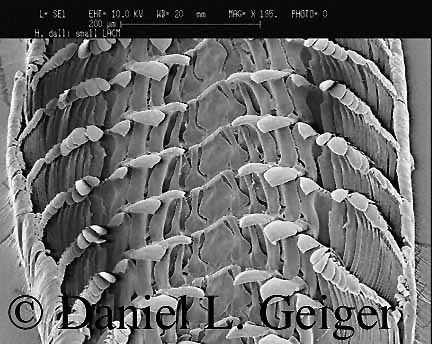
Above you see the full width of the radula of Haliotis dalli
Henderson, 1915, from the Galapagos Islands. The radula in this species
has very narrow laterals 1 as you will be able to compare in the picture
below, showing some additional species. Otherwise you can clearly see,
that the two sides of the radula from a zipper, i.e., the teeth of one
half of the radula fold neatly in the space between two rows on the
other side. Back to top of page

Central Field: Rachidian
and Lateral Tooth 1
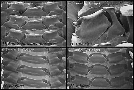
In these four sample pictures you can note a number of differences between
the species. In the top left corner Haliotis kamtschatkana has
the normal width of the rachidian tooth which does not have a basal
projection as in H. roberti to the right. H. elegans
has an extremely wide rachidian tooth, however, this character state
is currently based on a single specimen of this rather uncommon species,
hence needs verification. Note also, that neither of the two species
at the bottom have a basal projection of the rachidian tooth.
On the lateral tooth 1 you
note in H. kamtschatkana a concave depression on the ridge which
is a continuation of the cutting edge of the rachidian and the lateral
tooth 1. This depression cannot be found in either of the two species
shown at the bottom of the picture; these are rather convex. As already
mentioned, H. dalli has a special condition, in that the lateral
tooth 1 is extremely narrow, a synapomorphy it shares with H. roberti
McLean, 1970, from the Cocos Island. Back to top of page

Lateral Teeth 3-5
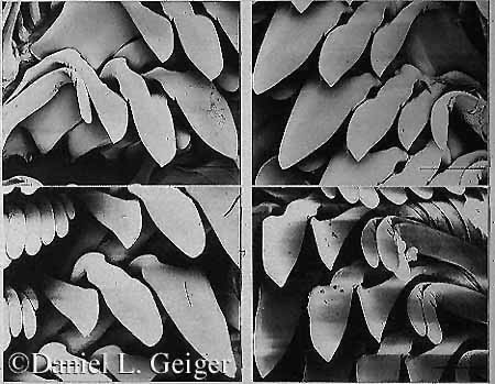
On the lateral teeth 3-5 only a single character could be extracted
so far: The presence and absence of denticles on the outer margin of
these teeth. I have illustrated this character with two pairs of examples.
In the top two panels the denticles are visible, whereas the lower two
panels do not show them. Back to top of page

Marginals
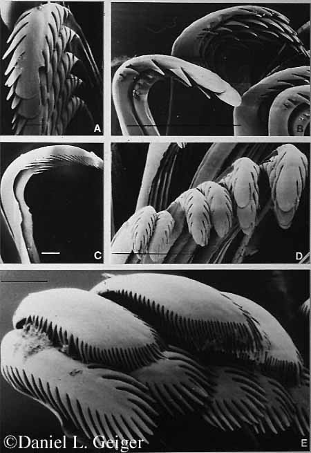
On the marginal teeth I found a single three-state character. In most
species the denticles are symmetrical, i.e., the first denticle on either
side of the cusp are on the same level as viewed on the longitudinal
axis of the cusp (panel A). In some species the denticles are off by
a count of one, and in a single species so far (Haliotis asinina
Linné, 1758) it is off by two: the first denticle on one side is
at the same level as the third on the other side (Panel D). Note, that
the number of denticles increased progressively while they become located
further from the middle of the radula. Panel C shows a intermediate
marginal in lateral view, and panel D shows outer marginals with very
fine brush shaped denticles even surrounding the tip of the cusp. Note
however, that the median denticle is a bit stronger developed into a
bristle. The outermost marginals have a further derived morphology not
illustrated here. Back to top of page
Conclusion
I hope I have successfully dispelled the myth, that the rhipidoglossan
radula of the Haliotidae is of no use for species level systematics.
So far only a dozen characters could be extracted from the radula, so
a phylogenetic analysis of the 55 species cannot be based upon the radula
alone. However, I has shown its usefulness as part in a total evidence
cladistic analysis. Back to top of page
Acknowledgments
I would like to acknowledge the support by the Labor für Rasterelektronenmikroskopie
of the University of Basel, where I could do some of the initial observations
on the intra- and inter-specific variability of the radula of abalone.
Further work in conjunction with my ongoing research for my dissertation
project at the University of Southern California was supported by a
Student Research Grants from the
Hawaiian Malacological Society and the Lehrner-Gray Fund for Marine
Research (American Museum of Natural History). Back to
top of page
| 


